What Excites One Mayo Clinic Radiologist About AI in Her Field
December 8, 2023
Source: drugdu
 393
393
Dr. Wendaline VanBuren, a radiology chair at Mayo Clinic, thinks that AI is in the beginning stages of improving radiologists’ workflows. Some of the most developed radiology AI research projects at Mayo center on image segmentation and 3D printing, she said. In the future, she’s excited to see more tools that aid radiologists in triage and lesion measurement.
By KATIE ADAMS

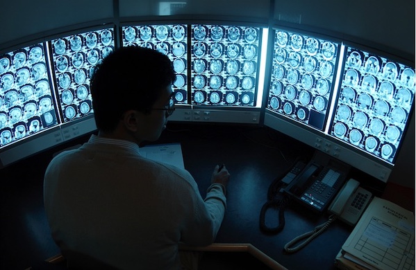
Radiology, like most physician specialties, is dealing with a labor shortage driven by burnout and an aging workforce. AI is often heralded as something that can help solve these workforce problems, but a lot of AI deployment in radiology is still in its iterative phase, said Dr. Wendaline VanBuren, chair of the gynecological imaging section within the division of abdominal radiology at Mayo Clinic.
Dr. VanBuren made this remark last week during an interview at RSNA 2023, the annual radiology and medical imaging conference in Chicago. While AI still has a long way to go before it makes a lasting impact on radiology’s workforce crisis, she said she thinks the technology has promising potential to change the branch of medicine for the better.
A lot of Mayo Clinic’s AI research centers around image segmentation, Dr. VanBuren noted. Segmentation involves dividing an image into meaningful regions, which is particularly useful for identifying and delineating structures or abnormalities. For example, clinicians could use these tools to segment organs, tumors or other structures in medical images to aid in diagnosis, treatment planning and disease monitoring.
Advancing segmentation research is exciting because good segmentation tools can help with 3D imaging, Dr. VanBuren declared.
Mayo Clinic’s exploration into the 3D image printing space is deepening, she said. 3D printing allows for the creation of physical, tangible models from medical imaging data. With these models, physicians and surgeons can gain a more intuitive and comprehensive understanding of complex anatomical structures. Clinicians can use these models to better educate patients, trainees and medical students, Dr. VanBuren noted.
“It’s an interesting development that 3D printing is already being employed clinically — that’s definitely an advancement in practice,” she remarked.
When looking toward the future of AI, Dr. VanBuren said she would like to see more workflow tools that help radiologists quickly access patient information. For instance, she said radiologists would benefit from an AI-assisted triage system that automatically organizes information regarding a patient’s medical background, indication for the exam and demographics.
In addition, Dr. VanBuren also called for AI tools to assist clinicians with the measurement of lesions. This task isn’t all that complex, but it’s pretty time-consuming, which means it could be an “easy target” for AI developers, she noted.
Say that a radiologist is following masses in their patients’ liver metastases. The radiologist must measure all of the lesions in three planes of imaging and then compare them to the previous exam in order to determine the response to treatment, Dr. VanBuren pointed out.
“Say we had an AI algorithm that could then compute that measurement aspect for us — that would really expedite the time. We could kind of just then do a visual inspection, and we wouldn’t have to do everything quantitatively,” she explained.
Photo: Hemera Technologies, Getty Images
Read more on
- The first subject has been dosed in the Phase I clinical trial of Yuandong Bio’s EP-0210 monoclonal antibody injection. February 10, 2026
- Clinical trial of recombinant herpes zoster ZFA01 adjuvant vaccine (CHO cells) approved February 10, 2026
- Heyu Pharmaceuticals’ FGFR4 inhibitor ipagoglottinib has received Fast Track designation from the FDA for the treatment of advanced HCC patients with FGF19 overexpression who have been treated with ICIs and mTKIs. February 10, 2026
- Sanofi’s “Rilzabrutinib” has been recognized as a Breakthrough Therapy in the United States and an Orphan Drug in Japan, and has applied for marketing approval in China. February 10, 2026
- Domestically developed blockbuster ADC approved for new indication February 10, 2026
your submission has already been received.
OK
Subscribe
Please enter a valid Email address!
Submit
The most relevant industry news & insight will be sent to you every two weeks.


