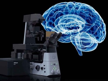
First-Of-Its-Kind AI-Enabled Smart Microscopy Platform to Accelerate Traditional Pathology Workflows
August 21, 2024
Source: drugdu
 278
278
 Pathology and tissue analysis are areas poised for transformative advancements. Drug developers and clinicians currently depend on long-established methods for crucial tasks such as diagnosing diseases, quantifying biomarkers, and predicting therapeutic responses. While there have been attempts to innovate by digitizing specimens and adding multiple markers to a single slide, there are still limitations, including the analysis of less than 1% of tissue samples and the inability to depict complex tissue architectures and cellular interactions that are only visible in three dimensions. 3D imaging technology captures significantly more data than traditional slide-based methods by digitizing whole biopsy specimens rather than just thin slices.Artificial intelligence (AI) and machine learning algorithms play a crucial role in quantifying relevant biomarkers and identifying areas for more detailed pathologist examination. Now, a pioneering 3D spatial biology platform can digitize entire tissue specimens quickly and non-destructively while providing AI-enabled quantitative analysis. This technology enhances the precision and confidence in addressing critical questions from clinicians and drug developers.
Alpenglow Biosciences (Seattle, WA, USA) is at the forefront of revolutionizing pathology with its patented multi-resolution 3Di platform for 3D spatial biology. In partnership with CorePlus, a prominent pathology lab (Puerto Rico) known for embracing AI-enabled digital pathology and Alpenglow's 3Di platform, they aim to elevate pathology from two-dimensional, qualitative analysis to three-dimensional, quantitative analysis. In a collaborative effort, CorePlus will annotate 3D prostate biopsy images to train AI models to identify critical tissue structures, such as cancer cells, immune cells, and blood vessels. Alpenglow will continue to refine its smart microscopy platform, which utilizes an innovative multi-resolution open top light sheet microscope. The current workflow for selecting regions of interest is manually conducted after data processing. Alpenglow plans to devise solutions for the rapid processing of large microscopy datasets, which can be a terabyte or larger, using AI to detect areas of interest across vast tissue volumes with higher sub-micron resolution for more thorough quantitative analysis.
To develop next-generation imaging technology powered by AI, Alpenglow is integrating NVIDIA Holoscan, a leading platform for developing medical devices, enabling AI edge computing on the 3Di system. Additionally, Alpenglow will employ NVIDIA IGX, an industrial-grade edge AI hardware platform, to enhance the performance of the 3Di. As a participant in the NVIDIA Inception program, which supports advanced startups, Alpenglow is closely collaborating with the NVIDIA healthcare team to optimize its GPU-accelerated image processing pipeline. This allows for rapid processing of extensive datasets and automated detection of significant regions within a tissue. Alpenglow has received a USD 1.6 million grant to automate the identification of regions within cancer biopsies that may require further analysis, potentially streamlining diagnoses and tailoring responses to novel treatments. This approach promises to drastically reduce the traditional pathology workflow time from days or weeks to mere hours, utilizing entire tissue samples for analysis.
"We are excited to continue to lead the field of 3D spatial biology and develop smart microscopy solutions to address cancer with this latest award," said Dr. Nicholas Reder, MD, MPH and CEO of Alpenglow Biosciences.
"Our team is thrilled to be advancing the transformation in pathology to incorporate 3D technology taking into consideration the inclusion of Hispanics to minimize racial bias in algorithm development," said Mariano de Socarraz, Founder and CEO at CorePlus. "Our collaboration with Alpenglow Biosciences to further develop rapid and high throughput AI-enabled 3D imaging of entire cancer biopsies will provide pathologists with unprecedented tools to improve precision in cancer diagnostics."
Source:
https://www.labmedica.com/pathology/articles/294802200/first-of-its-kind-ai-enabled-smart-microscopy-platform-to-accelerate-traditional-pathology-workflows.html
Read more on
- The first subject has been dosed in the Phase I clinical trial of Yuandong Bio’s EP-0210 monoclonal antibody injection. February 10, 2026
- Clinical trial of recombinant herpes zoster ZFA01 adjuvant vaccine (CHO cells) approved February 10, 2026
- Heyu Pharmaceuticals’ FGFR4 inhibitor ipagoglottinib has received Fast Track designation from the FDA for the treatment of advanced HCC patients with FGF19 overexpression who have been treated with ICIs and mTKIs. February 10, 2026
- Sanofi’s “Rilzabrutinib” has been recognized as a Breakthrough Therapy in the United States and an Orphan Drug in Japan, and has applied for marketing approval in China. February 10, 2026
- Domestically developed blockbuster ADC approved for new indication February 10, 2026
your submission has already been received.
OK
Subscribe
Please enter a valid Email address!
Submit
The most relevant industry news & insight will be sent to you every two weeks.


