AI Model Identifies Breast Tumor Stages Likely To Progress to Invasive Cancer
July 26, 2024
Source: https://www.labmedica.com/pathology/articles/294801919/ai-model-identifies-breast-tumor-stages-likely-to-progress-to-invasive-cancer.html
 361
361
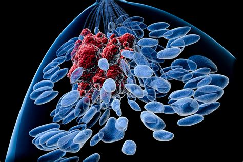
Ductal carcinoma in situ (DCIS) is a non-invasive type of tumor that can sometimes progress to a more lethal form of breast cancer and represents about 25% of all breast cancer cases. Between 30% and 50% of DCIS patients may develop an invasive stage of cancer, yet identifying which tumors will progress is still a challenge due to unknown biomarkers. Current diagnostic practices include multiplexed staining or single-cell RNA sequencing to determine DCIS stages in tissue samples, but these methods are costly and not widely used. This has led to potential overtreatment of patients with DCIS. Now, a new artificial intelligence (AI) model can distinguish different stages of DCIS from inexpensive and readily available breast tissue images.
The model developed by an interdisciplinary team of researchers from MIT (Cambridge, MA, USA) and ETH Zurich (Zurich, Switzerland) was trained and tested using one of the largest datasets of its kind that built because such tissue images are so easy to obtain. This AI model could potentially streamline the diagnosis process for simpler DCIS cases, reducing reliance on labor-intensive methods and allowing clinicians to focus more on ambiguous cases. Previously, the team found that a low-cost imaging technique called chromatin staining could deliver insights comparable to those from high-cost single-cell RNA sequencing. They hypothesized that combining this staining method with a sophisticated machine-learning model could yield detailed cancer stage information at a lower cost.
They compiled a dataset of 560 tissue sample images from 122 patients across three disease stages to train their AI model. This model learns to represent the state of each cell within an image to determine the cancer's stage. Recognizing that not all cells indicate cancer presence, the team engineered the model to create clusters of cells with similar states, identifying eight distinct states critical for diagnosing DCIS. Some states suggest a higher likelihood of invasive cancer. However, they learnt that knowing the proportion of each cell state was insufficient; understanding how these cells are organized within the tissue was also crucial. The model was enhanced to assess both the proportion and spatial arrangement of cell states, thereby significantly improving its accuracy. When compared to traditional pathologist evaluations, the model showed high concordance in many cases. For less definitive cases, the model provided insights into tissue sample features, like cell organization, which could aid pathologists in their diagnostics. This model’s versatility suggests potential applications beyond breast cancer to other cancers and neurodegenerative diseases, areas the researchers are currently exploring.
“We took the first step in understanding that we should be looking at the spatial organization of cells when diagnosing DCIS, and now we have developed a technique that is scalable,” said MIT’s Caroline Uhler. “From here, we really need a prospective study. Working with a hospital and getting this all the way to the clinic will be an important step forward.”
By editor
Copyright©2026 Ddu. All rights reserved.
Read more on
- Chinese innovative drugs score a Blockbuster Deal! AstraZeneca buys out Suvolotinib for $1.5 billion July 15, 2026
- 【Company Recommendation】Shanghai Wantewant Technology Co., Ltd July 10, 2026
- Ascletis Pharma submits Investigational New Drug (IND) applications to the FDA for two new obesity drugs July 9, 2026
- Fengtou News | Highlight Pharmaceuticals’ Ganoxitinib Receives Breakthrough Therapy Designation from CDE July 9, 2026
- 【Company Recommendation】Emeishan Hongsen Biopharmaceutical Co., Ltd. June 26, 2026
your submission has already been received.
OK
Subscribe
Please enter a valid Email address!
Submit
The most relevant industry news & insight will be sent to you every two weeks.