COPD Assessment by CT Analysis
July 2, 2018
Source: IndiaTimes
 2,443
2,443
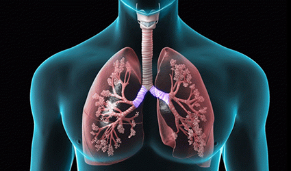
Chronic Obstructive Pulmonary Disease (COPD) is defined as a condition which affects airways and alveoli, leading to the loss of lung function in a progressive manner. COPD is the fourth leading cause of death globally and has affected more than 200 million people throughout the world.
A research team from the University of Southampton in the UK have developed a novel method to analyze CT images of lungs for an easy assessment and diagnosis of COPD, which was reported in the journal, Scientific Reports. By combining computed tomography (CT) scans and high-performance computing algorithms, the numerical analysis of the entire lungs can be assessed. Features such as size and structure of bronchial parts, direction of the bronchial branches and the changes in lung shapes can be studied during deep inspiration and expiration. A larger, complex bronchial portion indicates good lung function whereas, a smaller distorted lung part shows poor lung function.
Early diagnosis followed by the severity of lung condition could be assessed by comparing its results across a larger database of images. Even lung density and bronchial wall thickness were also used to examine and interpret CT lung images.
Jacek Brodzki, a professor at the University of Southampton said, "Our study shows that this new method, employing topological data analysis, can complement and expand on established techniques to give a valuable, accurate range of information about the lung function of individuals."
Ratko Djukanovic, a professor at the University of Southampton said, "This method is a major advance in our ability to study the structural abnormalities of COPD, a complex disease that affects so many people and, sadly, results in significant morbidity and mortality."
By Ddu
Copyright©2026 Ddu. All rights reserved.
Read more on
- 【EXPERT Q&A】What are the regulations and requirements for exporting medical devices to the European Union? September 5, 2023
- 【EXPERT Q&A】What is the procedure for registering medical devices for the Russian market? August 22, 2023
- Things to Know before Buying Newborn Baby Incubators March 31, 2022
- Portable Nebulizer Machine September 10, 2018
- PHYSIOTHERAPY TABLE September 7, 2018
your submission has already been received.
OK
Subscribe
Please enter a valid Email address!
Submit
The most relevant industry news & insight will be sent to you every two weeks.