New 3D Color X-Rays to Diagnose Cancer, Heart Disease and More
July 20, 2018
Source: Medical Design and Outsourcing
 1,876
1,876
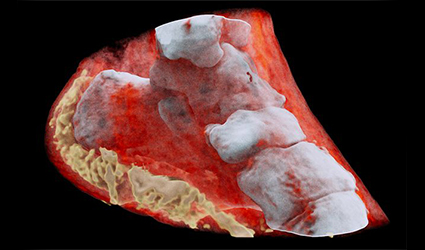
The first-ever 3D, color X-rays were tested on a human by New Zealand Scientists, using technology that promises to enhance medical diagnostics in oncology, cardiology, neurology and orthopedics.
The scanner incorporates the Medipix3RX detector chip, a particle-tracking technology built for the CERN Large Hadron Collider, based on the traditional black-and-white X-ray technology, which was a product of the Medipix3 Collaboration, comprising CERN in Geneva and 18 other research institutions worldwide.
Phil Butler, a physics professor at the University of Canterbury in Christchurch, N.Z. said, “The machine’s small pixels and accurate energy resolution meant that this new imaging tool is able to get images that no other imaging tool can achieve,”
Researchers were able to detect varied cell lines in cancerous tissue in preclinical trials on animals at universities in New Zealand, Europe and the United States, Anthony Butler said in a phone interview. The scanner could also measure arterial plaque non-invasively thereby identifying the risk of heart attack or stroke in patients and track their progress post-treatment, he added.
Bone diseases such as arthritis and osteoporosis can also be studied using this scanner. In the coming months, a clinical trial of orthopedic and rheumatology patients is planned in New Zealand. The CERN says that the images not only very clearly differentiate between bone, muscle and cartilage, but also show the position and size of cancerous tumors.
“This color X-ray imaging technique could produce clearer and more accurate pictures and help doctors give their patients more accurate diagnoses,” said a CERN statement.
By Ddu
Copyright©2026 Ddu. All rights reserved.
Read more on
- 【EXPERT Q&A】What are the regulations and requirements for exporting medical devices to the European Union? September 5, 2023
- 【EXPERT Q&A】What is the procedure for registering medical devices for the Russian market? August 22, 2023
- Things to Know before Buying Newborn Baby Incubators March 31, 2022
- Portable Nebulizer Machine September 10, 2018
- PHYSIOTHERAPY TABLE September 7, 2018
your submission has already been received.
OK
Subscribe
Please enter a valid Email address!
Submit
The most relevant industry news & insight will be sent to you every two weeks.