3D Pathology with AI to Enhance Prognosis Accuracy for Barrett’s Esophagus Patients
August 10, 2024
Source: drugdu
 279
279
 Barrett's esophagus is a condition where the lining of the esophagus changes due to chronic gastroesophageal reflux. Individuals with Barrett's esophagus are at a slightly increased risk of developing esophageal cancer and require regular surveillance endoscopies. During these procedures, gastroenterologists collect numerous biopsies from the affected tissues. These samples are then cut into thin sections and placed on glass slides for examination under a microscope by pathologists. However, the tissue sections that pathologists view represent only about 1% or less of the actual biopsies and provide just a two-dimensional view, which can be misleading. Researchers are now conducting clinical studies of archived tissues from patients with the condition to develop computational 3D pathology methods for Barrett’s esophagus risk stratification.
The research team at UW College of Engineering (Seattle, WA, USA) had previously invented 3D pathology methods to assess prostate cancer risk and shifted their focus on gastrointestinal applications of their technologies, including for evaluating esophageal cancer risk in patients with Barrett’s esophagus. They aim to demonstrate that analyzing 3D pathology datasets from entire endoscopic biopsies using AI can better determine which patients might progress to esophageal cancer and thus require more intensive treatment. The team is utilizing open-top light-sheet microscopy for this purpose. This innovative technique allows for 3D microscopic viewing of biopsies without the need for slicing, preserving the entire tissue structure.
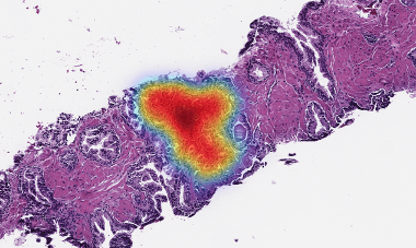
This "slide-free" microscopy technique involves using a light sheet and high-speed cameras to image tissue samples stained with fluorescent dyes and made transparent through a process called optical clearing. Once the 3D pathology datasets are prepared, AI is employed to either highlight the most crucial areas of the biopsy for pathologist review or to autonomously evaluate the tissues. In previous research published in Modern Pathology, the team introduced a deep learning approach that proved more efficient at identifying malignancies in Barrett’s esophagus biopsies than traditional methods, significantly reducing the number of images pathologists need to examine. Furthermore, the team is enhancing the AI model's training process by developing an advanced weakly-supervised deep learning triage system for analyzing 3D pathology datasets.
“We are trying to identify the highest risk patients so that they may receive early treatments that could be critical for their survival,” said Professor Jonathan Liu, professor of mechanical engineering, bioengineering, and laboratory medicine & pathology at the University of Washington. “In our archived tissue samples, some patients progressed to cancer, and we are trying to detect what in their tissues could have predicted that at the earliest stages.”
Source:
https://www.labmedica.com/pathology/articles/294802116/3d-pathology-with-ai-to-enhance-prognosis-accuracy-for-barretts-esophagus-patients.html
Read more on
- The first subject has been dosed in the Phase I clinical trial of Yuandong Bio’s EP-0210 monoclonal antibody injection. February 10, 2026
- Clinical trial of recombinant herpes zoster ZFA01 adjuvant vaccine (CHO cells) approved February 10, 2026
- Heyu Pharmaceuticals’ FGFR4 inhibitor ipagoglottinib has received Fast Track designation from the FDA for the treatment of advanced HCC patients with FGF19 overexpression who have been treated with ICIs and mTKIs. February 10, 2026
- Sanofi’s “Rilzabrutinib” has been recognized as a Breakthrough Therapy in the United States and an Orphan Drug in Japan, and has applied for marketing approval in China. February 10, 2026
- Domestically developed blockbuster ADC approved for new indication February 10, 2026
your submission has already been received.
OK
Subscribe
Please enter a valid Email address!
Submit
The most relevant industry news & insight will be sent to you every two weeks.


